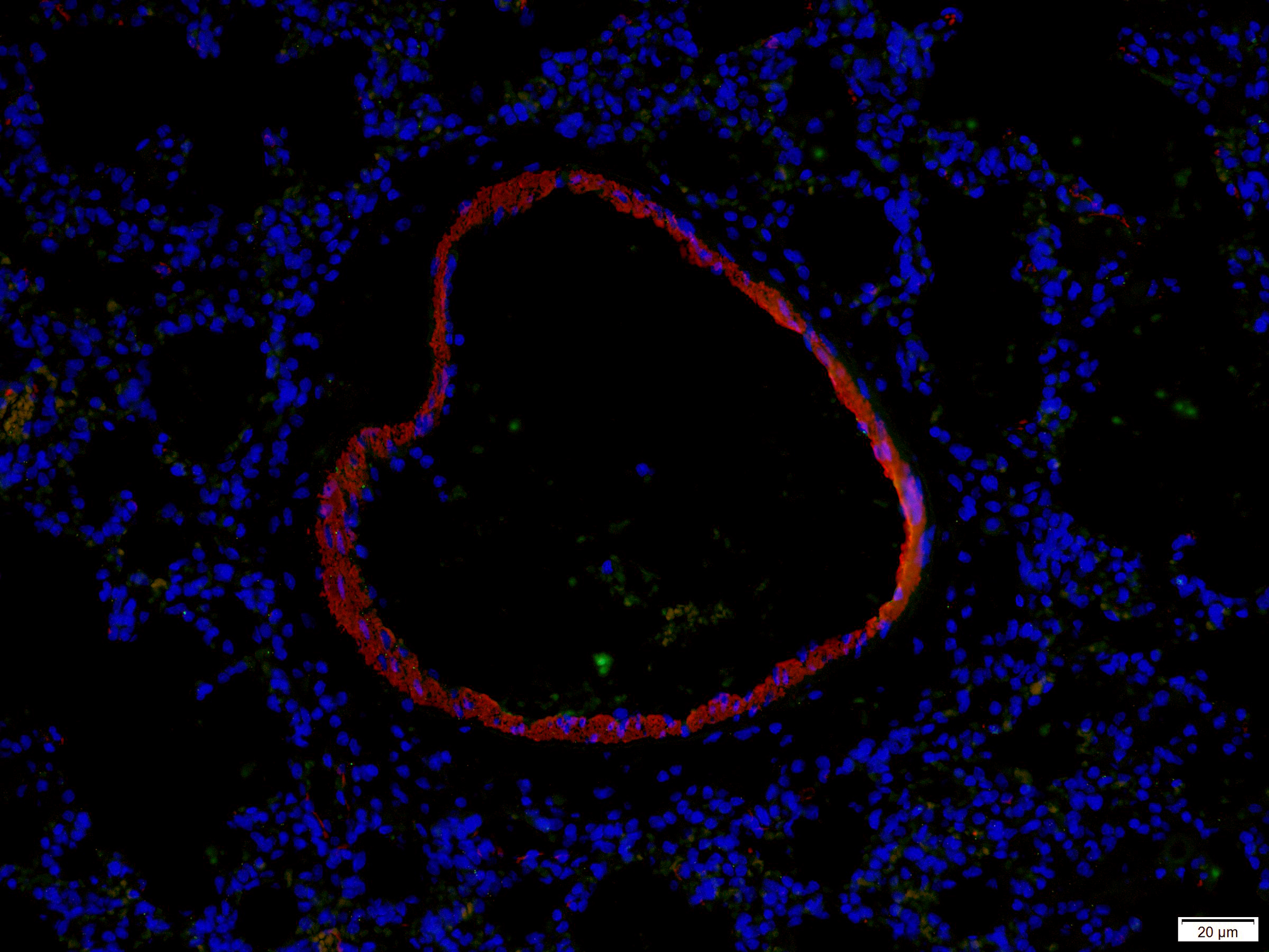
Polink-2 Plus HRP Polymer Detection for MOUSE and RABBIT antibody on human tissue
Products
Polink-2 Plus HRP Polymer Detection for MOUSE and RABBIT antibody on human tissue
Polink-1 HRP Polymer Detection for RABBIT antibody on human or animal tissue
Citrate Buffer Solution pH 6.0 (20x)
Polink-1 HRP Polymer Detection for RAT antibody on human or animal tissue
AEC concentrated Kit (20x) for 2000 slides
Polink-2 Plus HRP Polymer Detection for RABBIT antibody on human or animal tissue
DAB Enhancer Concentrate (20x)
Polink-2 Plus HRP Polymer Detection for RABBIT antibody on human or animal tissue
Aqueous mounting medium for preserving fluorescence of tissue and cell smears
Polink-1 HRP Polymer Detection for MOUSE antibody on human or animal tissue (non-rodent)
Polink-2 Plus HRP Polymer Detection for MOUSE antibody on human or animal tissue (non-rodent)
Polink-2 Plus AP Polymer Detection for MOUSE antibody on human or animal tissue (non-rodent)
Pepsin Kit inlcudes buffer (RTU)
Aqueous mounting medium for preserving fluorescence of tissue and cell smears. This mounting medium is fortified with DAPI which is a counterstain for DNA.

![IHC staining of FFPE human breast cancer tissue using mouse anti-ER polyclonal antibody and Polink-2 Plus HRP Broad for DAB detection kit D41-18. The brown stain indicates positive stain and blue is the counter stain. It is mounted using a permanent organic mounting medium [E37-100].](https://cdn.origene.com/catalog/product/assets/images/assay-kit/ihc-kit-and-reagent/151/d41.jpg?browse)


![Human tonsil with rabbit anti FoxP1 (clone EP137) using Polink1 (D13-18) rabbit polymer detection and HIER Accel ([B22C-125])for was used 10 minutes pressure cooker. Primary antibody was incubated 1 hour; Polink-1 (D13-18) for 15 minutes and chromogen for 5 minutes according to protocol data sheet.](https://cdn.origene.com/catalog/product/assets/images/assay-kit/ihc-kit-and-reagent/151/d13-18-102.jpg?browse)
![Human tonsil with rabbit anti FoxP1 (clone EP137) using Polink1 (D13-18) rabbit polymer detection and HIER Accel ([B22C-125])for was used 10 minutes pressure cooker. Primary antibody was incubated 1 hour; Polink-1 (D13-18) for 15 minutes and chromogen for 5 minutes according to protocol data sheet.](https://cdn.origene.com/catalog/product/assets/images/assay-kit/ihc-kit-and-reagent/151/d13-18-1.jpg?browse)


![IHC staining of FFPE human skin tissue within normal limits using rat anti-podoplanin polyclonal antibody and Polink-1 HRP Rat NM for DAB detection kit D35-18. The brown stain indicates positive stain and blue is the counter stain. It is mounted using a permanent organic mounting medium [E37-100].](https://cdn.origene.com/catalog/product/assets/images/assay-kit/ihc-kit-and-reagent/151/d35.jpg?browse)
![Mouse heart muscle screened with Rat anti HSF1 (SantaCruz; 1:100). HIER buffer TEE ([B21-100]) was used in pressure cooker. Secondary Polink 1 HRP Rat with DAB Chromogen (D35-18) was use according to protocol data sheet.](https://cdn.origene.com/catalog/product/assets/images/assay-kit/ihc-kit-and-reagent/151/d35-18-100-h.jpg?browse)
![Mouse testis muscle screened with Rat anti HSF1 (SantaCruz; 1:100). HIER buffer TEE ([B21-100]) was used in pressure cooker. Secondary Polink 1 HRP Rat with DAB Chromogen (D35-18) was use according to protocol data sheet.](https://cdn.origene.com/catalog/product/assets/images/assay-kit/ihc-kit-and-reagent/151/d35-18-101-h.jpg?browse)

![IHC staining of FFPE human tonsil tissue within normal limits using mouse anti-human B cell polyclonal antibody, Polink-1 HRP Mouse for AEC detection kit [D15-18], and AEC Kit (Cat. No.: C01-12). The red stain indicates positive stain. It is mounted using a permanent aqueous mounting medium [E03-18].](https://cdn.origene.com/catalog/product/assets/images/assay-kit/ihc-kit-and-reagent/151/c01.jpg?browse)
![Human tonsil stained with with OriGene Mouse anti Ki67 ([UM800033] diluted 1:2000) seen as the red brick nuclear stain HIER buffer TEE ([B21C-100]) was used 10 minutes pressure cooker. Secondary SPlinked anti-Ms & Rb ([D01-18]), AEC chromogen (C01-12) was used according to data sheet instructions](https://cdn.origene.com/catalog/product/assets/images/assay-kit/ihc-kit-and-reagent/151/c01-12-1.jpg?browse)
![IHC staining of FFPE human pancreas tissue within normal limits using rabbit anti-glucagon polyclonal antibody and Polink-2 Plus HRP Rabbit for DAB detection kit [D39-18]. The brown stain indicates positive stain and blue is the counter stain. It is mounted using a permanent organic mounting medium [E37-100].](https://cdn.origene.com/catalog/product/assets/images/assay-kit/ihc-kit-and-reagent/151/d39.jpg?browse)

![IHC staining of FFPE human pancreas tissue within normal limits using rabbit anti-glucagon polyclonal antibody, Polink-1 HRP Rabbit for DAB detection kit [D13-18], and DAB Enhancer Kit (Cat. No.: C06). The brown stain indicates positive stain and blue is the counter stain. It is mounted using a permanent organic mounting medium [E37-100].](https://cdn.origene.com/catalog/product/assets/images/assay-kit/ihc-kit-and-reagent/151/c06.jpg?browse)
![Human placenta stained with with OriGene Mouse anti PDL1 ([UM800120] diluted 1:100) seen as the brown left panel and black/brown on right Panel in the membrane and cytoplasmic. The stain was produced using HIER buffer TEE ([B21C-100]) and secondary SPlinked anti-Ms & Rb ([D01-18]), DAB chromogen ([C09-12]) only on left panel and DAB chromogen ([C09-12]) with DAB enhancer (C07-25) on right panel.](https://cdn.origene.com/catalog/product/assets/images/assay-kit/ihc-kit-and-reagent/151/c07-25-1.jpg?browse)
![Human tonsil stained with with OriGene Mouse anti-Ki67 ([UM800033] diluted 1:2000) seen as the brown left panel and black/brown on right Panel in the nuclear. The stain was produced using HIER buffer TEE ([B21C-100]) and secondary SPlinked anti-Ms & Rb ([D01-18]), DAB chromogen ([C09-12]) only on left panel and DAB chromogen ([C09-12]) with DAB enhancer (C07-25) on right panel.](https://cdn.origene.com/catalog/product/assets/images/assay-kit/ihc-kit-and-reagent/151/c07-25-102.jpg?browse)


![Human placenta with OriGene Mouse anti-PD-L1 ([UM800120] dilution 1:100) using Polink1 (D12-110) mouse polymer detection and HIER TEE buffer pH9 for was used 10 minutes pressure cooker. Primary antibody was incubated 1 hour; Polink-1 (D12-110) for 15 minutes and chromogen for 5 minutes according to protocol data sheet.](https://cdn.origene.com/catalog/product/assets/images/assay-kit/ihc-kit-and-reagent/151/d12-110-101.jpg?browse)
![Human placenta with OriGene Mouse anti-PD-L1 ([UM800120] dilution 1:100) using Polink1 (D12-110) mouse polymer detection and HIER TEE buffer pH9 for was used 10 minutes pressure cooker. Primary antibody was incubated 1 hour; Polink-1 (D12-110) for 15 minutes and chromogen for 5 minutes according to protocol data sheet.](https://cdn.origene.com/catalog/product/assets/images/assay-kit/ihc-kit-and-reagent/151/d12-110-1.jpg?browse)

![IHC staining of FFPE human tonsil tissue within normal limits using mouse anti-human B cell polyclonal antibody and Polink-2 Plus HRP Mouse for DAB detection kit [D37-18]. The brown stain indicates positive stain and blue is the counter stain. It is mounted using a permanent organic mounting medium [E37-100].](https://cdn.origene.com/catalog/product/assets/images/assay-kit/ihc-kit-and-reagent/151/d37.jpg?browse)

![Human colon cancer screened with OriGene Ms anti CEA ([TA803456] @ 1:500). HIER buffer Citrate ([B05-100B]) was used 10 minutes pressure cooker. Secondary Polink 2 plus AP Ms (D69-110) was use according to protocol data sheet. Cytoplasmic and membrane stain was visualized with GBI permanent red chromogen ([C13-120]).](https://cdn.origene.com/catalog/product/assets/images/assay-kit/ihc-kit-and-reagent/151/d69-110-1.jpg?browse)
![Human tonsil screened with OriGene Ms anti Ki67 ([UM800033] @ 1:1000). HIER buffer Citrate ([B05-100B]) was used 10 minutes pressure cooker. Secondary Polink 2 plus AP Ms (D69-110) was use according to protocol data sheet. Nuclear stain was visualized with GBI permanent red chromogen ([C13-120])](https://cdn.origene.com/catalog/product/assets/images/assay-kit/ihc-kit-and-reagent/151/d69-110-101.jpg?browse)
![Human colon cancer screened with OriGene Ms anti MS4A1 ([UM800009] @ 1:800). HIER buffer Citrate ([B05-100B]) was used 10 minutes pressure cooker. Secondary Polink 2 plus AP Ms (D69-110) was use according to protocol data sheet. Cytoplasmic and membrane stain was visualized with GBI permanent red chromogen ([C13-120]).](https://cdn.origene.com/catalog/product/assets/images/assay-kit/ihc-kit-and-reagent/151/d69-110-102.jpg?browse)

![IHC staining of FFPE human skin tissue within normal limits using mouse anti-cytokeratin monoclonal antibody, SPlink HRP Mouse for DAB detection kit [D02-18], and Pepsoin Solution (Cat. No.: E06-50). The brown stain indicates positive stain and blue is the counter stain. It is mounted using a permanent organic mounting medium [E37-100].](https://cdn.origene.com/catalog/product/assets/images/assay-kit/ihc-kit-and-reagent/151/e06.jpg?browse)


