More Specific Antibodies for Cancer Diagnostics
Case Studies Using High-Density Protein Microarray
Case Study 1: HER2 (also called ERBB2) Pathology Antibodies
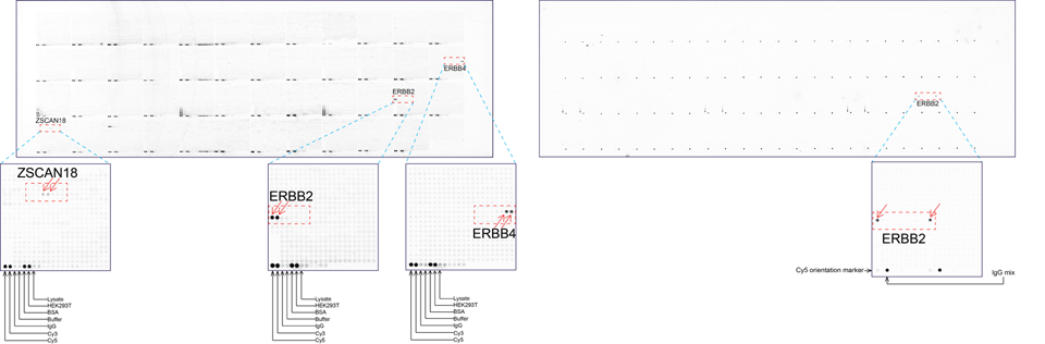
A semi-quantitative immunohistochemical assay using anti-HER2 antibody is applied to determine HER2 protein overexpression in breast cancer tissues. The specificity of the HER2 antibody (e.g. Clone4B5) is critical because the test results will help oncologist decide a patient should receive Herceptin™ treatment. By using OriGene’s 10K high-density protein microarray, we have revealed that the antibody 4B5 is not specific to HER2 protein. As shown in the graph above, this antibody also reacts with ZSCAN1B and HER4 (ERBB4). In contrast, HER2 UltraMAB (Clone UMAB36) developed by OriGene only recognizes HER2 protein and is thus specific.
Case Study 2: ERCC1 IHC antibodies
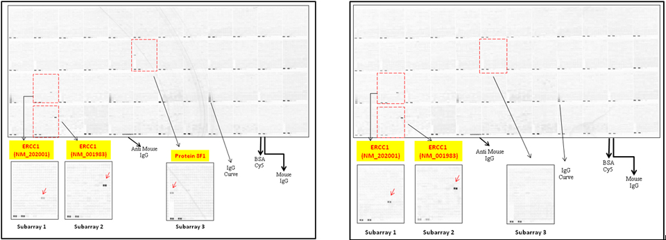
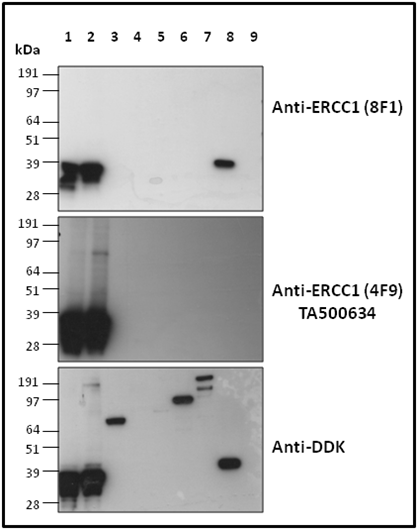
The excision repair cross-complementation group 1 (ERCC1) protein is an important biomarker for clinicians to predict whether certain patient populations with non-small cell lung carcinoma will respond to cisplatin chemotherapy. As such, it is critical to develop highly-specific immunohistochemistry validated monoclonal antibodies for this diagnostic test. Several publications revealed that 8F1, a commonly used antibody clone for ERCC1, exhibits cross-reactivity to an unknown protein in ERCC1 deficient cell lines. By using OriGene’s high-density protein microarray technology, the corresponding cross-reactive binding protein for 8F1 monoclonal antibody was identified. This technology also enable us successfully develop the most specific UltraMAB® monoclonal antibody for ERCC1 (4F9, Clone UMAB8). This data was further confirmed by western blot analysis. Review the publication on the validation of ERCC1 antibodies
| 抗体相关资料 |

